
Introduction And Epidemiology
- Kawasaki disease presenting with partial clinical criteria.
- Defined by unexplained fever lasting five or more days associated with only two or three classic clinical features.
- Highly prevalent in young infants under six months of age.
- Infant presenting with unexplained fever for seven or more days without apparent focus mandates immediate echocardiographic evaluation.
- Demonstrates profoundly higher risk for coronary artery abnormalities secondary to delayed clinical recognition.
- Clinical features frequently evolute sequentially over days, obscuring timely diagnosis.
Atypical Clinical Manifestations
- Standard mucocutaneous signs remain frequently absent, subtle, or fleeting.
- Reactivation of bacillus Calmette-Guerin inoculation site exhibits profound erythema and induration.
- Extreme irritability acts as prominent clinical clue, occurring secondary to aseptic meningitis.
- Node-first presentation manifests as isolated, unilateral, tender cervical lymphadenopathy.
- Neck ultrasonography reveals uniformly hypoechoic nodes lacking necrosis, demonstrating well-circumscribed margins termed cluster of grapes appearance.
- Acute hydrocele presents as sudden, painful unilateral scrotal swelling.
- Hydrops of gallbladder presents as upper abdominal mass, mimicking acute surgical abdomen.
- Perianal erythema with profound desquamation occurs frequently during acute disease phase.
Diagnostic Evaluation Algorithm
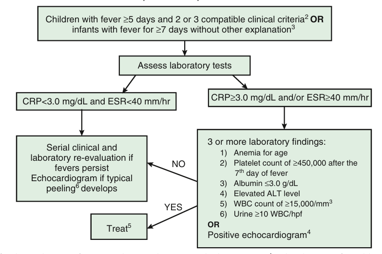
- Lacks pathognomonic diagnostic laboratory test.
- Evaluation strictly triggered when C-reactive protein equals or exceeds 3.0 mg/dL, or erythrocyte sedimentation rate equals or exceeds 40 mm/hr.
- Diagnosis established utilizing standardized diagnostic algorithms integrating supplemental laboratory and echocardiographic parameters.
Supplemental Laboratory Criteria
- Presence of three or more supplemental laboratory criteria establishes positive diagnostic threshold.
| Biomarker | Diagnostic Threshold |
|---|---|
| Hemoglobin | Anemia for age. |
| Platelet Count | Greater than or equal to 450,000 per cubic millimeter after seventh day of fever. |
| Serum Albumin | Less than or equal to 3.0 g/dL. |
| Hepatic Enzymes | Elevated alanine aminotransferase. |
| Leukocytes | White blood cell count greater than or equal to 15,000 per cubic millimeter. |
| Urinalysis | Greater than or equal to 10 white blood cells per high-power field indicating sterile pyuria. |
Echocardiographic Diagnostic Criteria
- Echocardiogram considered positive for incomplete disease if specific structural or functional conditions met.
| Finding Category | Specific Diagnostic Conditions |
|---|---|
| Coronary Dilation | Z score of left anterior descending or right coronary artery equals or exceeds 2.5. |
| Direct Observation | Coronary artery aneurysm directly visualized. |
| Suggestive Features | Presence of three or more subtle criteria establishes positive scan. Includes perivascular brightness, lack of vessel tapering. Includes decreased left ventricular function, mitral regurgitation, pericardial effusion. Includes Z scores in left anterior descending or right coronary artery between 2.0 and 2.5. |
Advanced Biomarkers
- N-terminal pro-B-type natriuretic peptide levels exceeding 500 pg/mL strongly assist diagnostic confirmation in ambiguous presentations.
- Gamma-glutamyl transferase elevation occurs notably without frank hepatic dysfunction.
Differential Diagnosis
- Presentation mimics multiple infectious, rheumatologic, and hyperinflammatory syndromes.
| Disease Category | Distinguishing Clinical And Laboratory Features |
|---|---|
| Multisystem Inflammatory Syndrome In Children | Occurs post-SARS-CoV-2 infection. Affects older demographic. Features profound gastrointestinal pain, severe myocardial dysfunction, profound lymphopenia, marked hyperferritinemia. |
| Systemic Juvenile Idiopathic Arthritis | Features quotidian fever pattern. Exhibits prominent hepatosplenomegaly. Lacks coronary artery aneurysms classically. |
| Adenovirus Infection | Demonstrates prominent exudative pharyngitis and exudative conjunctivitis. |
| Scarlet Fever | Yields rapid clinical response to appropriate antibiotic therapy. Demonstrates elevated anti-streptolysin O titers and sandpaper rash. |
| Macrophage Activation Syndrome | Features unremitting fever, profound hepatosplenomegaly. Laboratory profile reveals paradoxically dropping erythrocyte sedimentation rate, extreme hyperferritinemia, hypofibrinogenemia. |
Management Strategies
- Mandates immediate therapeutic intervention to halt progressive coronary architecture damage.
- Intravenous immunoglobulin 2 g/kg administered as single continuous infusion over 8 to 12 hours.
- Aspirin therapy initiated concurrently.
- Recent 2024 guidelines advocate low-dose aspirin 3 to 5 mg/kg/day from onset to minimize gastrointestinal bleeding risks.
- Antiplatelet aspirin continued minimum 6 to 8 weeks.
- Discontinue aspirin entirely if serial echocardiograms remain completely normal.
- Live viral vaccines deferred for 11 months following intravenous immunoglobulin administration.
Intensification And Refractory Therapy
- High-risk patients require immediate intensification of primary regimen.
- Infants under six months uniformly require intensification due to exceptional aneurysm risk approaching 43%.
- Baseline coronary artery Z score exceeding 2.5 strictly mandates intensification.
- Corticosteroids utilize intravenous methylprednisolone 2 mg/kg/day divided every 12 hours.
- Infliximab 10 mg/kg infused over 2 hours serves as potent biological alternative.
- Intravenous immunoglobulin resistance defined strictly as persistent or recrudescent fever 36 hours post-infusion.
- Resistance necessitates prompt administration of second intravenous immunoglobulin dose, pulse corticosteroids, or calcineurin inhibitors including cyclosporine.
- Statins (75 mg/kg/day) modulate endothelial function, block endothelial to mesenchymal transition, and reduce oxidative stress in persistent disease.
Complications And Prognosis
- Delayed clinical diagnosis independently drives highest rates of coronary artery aneurysms in infants.
- Giant aneurysms (Z score greater than or equal to 10) carry lifetime risk of occlusive thrombosis, myocardial infarction, and sudden death.
- Giant aneurysms necessitate lifelong dual antiplatelet therapy coupled with systemic anticoagulation utilizing low molecular weight heparin or direct oral anticoagulants.
- Myocardial infarction risk peaks dramatically during first 2 to 3 months following illness onset.
- Complete regression of small aneurysms occurs frequently, carrying near-zero long-term risk of adverse cardiac events.
- Regressed large aneurysms maintain permanent myointimal thickening and abnormal vascular reactivity indefinitely.
- Mandatory long-term surveillance incorporates advanced imaging modalities including computed tomography angiography or magnetic resonance imaging to detect progressive luminal stenosis.