
Definition And Core Concepts
- Laboratory technique for visualization and analysis of chromosomes in metaphase cells to determine number, size, shape, and banding pattern.
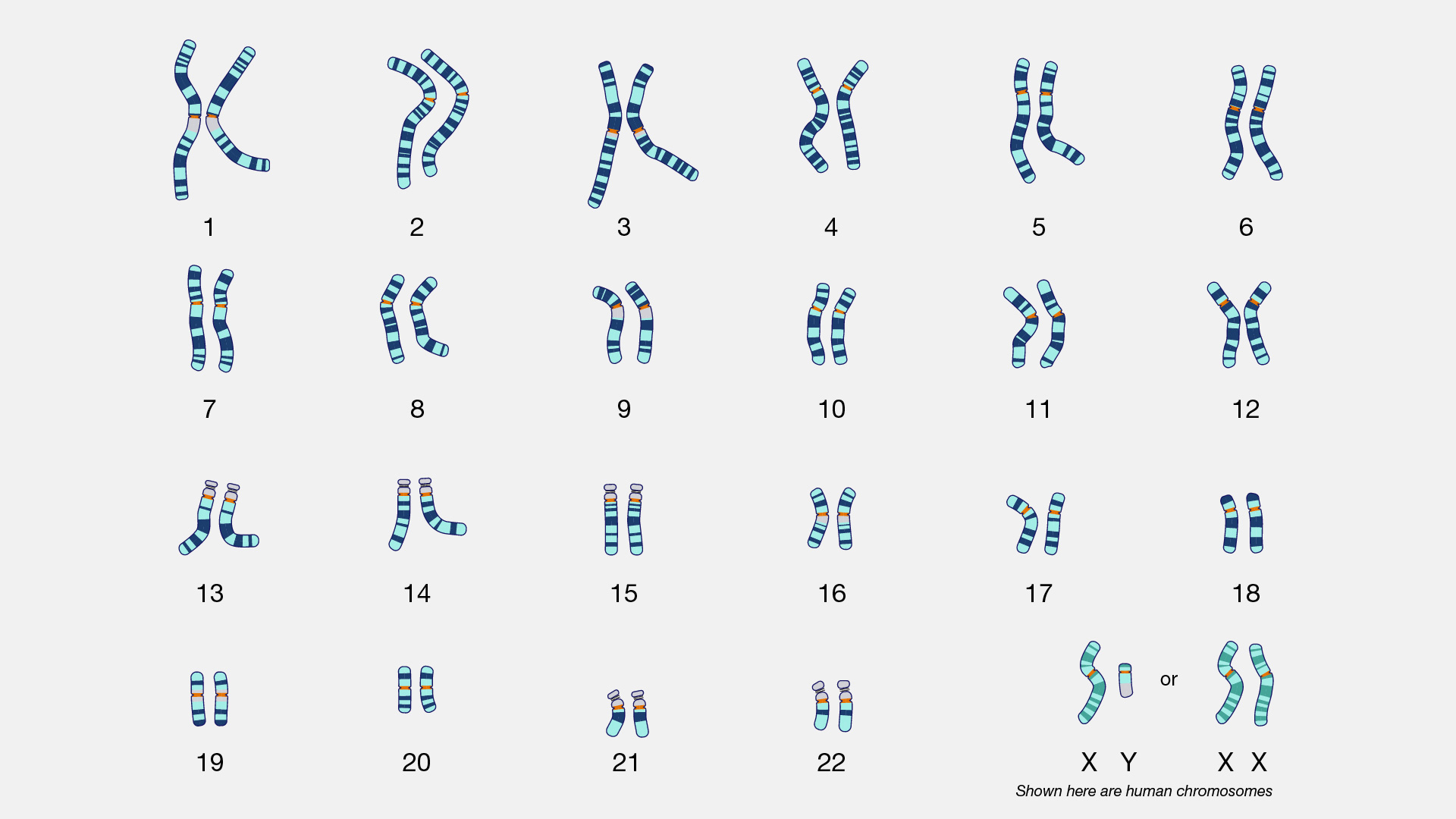
- Produces a karyogram, a digital representation organizing the entire genome's paired chromosomes.
- Chromosomes are classified based on length (chromosome 1 is longest, 22 is smallest), banding pattern, and centromere position (metacentric, submetacentric, or acrocentric).
- Standard resolution is 5-10 Megabases (Mb), allowing detection of numerical abnormalities (aneuploidies) and large structural rearrangements.
Methodology And Procedure
- Sample Collection: Requires actively dividing cells sourced from peripheral blood lymphocytes, skin fibroblasts, bone marrow aspirates, chorionic villi, or amniotic fluid.
- Cell Culture: Phytohemagglutinin (PHA) stimulation is utilized for T-lymphocytes over 72 hours.
- Mitotic Arrest: Colchicine or colcemid is added to block spindle formation, arresting cells in metaphase or prometaphase.
- Hypotonic Treatment: Swells cells and disrupts the nuclear membrane, facilitating proper dispersion of chromosomes on the slide.
- Fixation: Utilizes a methanol and acetic acid mixture.
- Banding And Staining: GTG banding (G bands by trypsin using Giemsa) is the most widely utilized method, producing a unique sequence of dark (G-positive) and light (G-negative) bands.
- Microscopic Analysis: Standard metaphase spread provides 450-550 bands. High-resolution analysis utilizes less-condensed prometaphase chromosomes, yielding 550-850 bands. A minimum of 20-30 metaphases are analyzed to detect mosaicism.
Clinical Indications
| Category | Specific Indications |
|---|---|
| Suspected Aneuploidy Syndromes | Clinical features of Down syndrome (Trisomy 21), Edwards syndrome (Trisomy 18), Patau syndrome (Trisomy 13), Turner syndrome (45,X), or Klinefelter syndrome (47,XXY). |
| Pediatric And Developmental | Multiple congenital anomalies, dysmorphic features, unexplained global developmental delay or intellectual disability. |
| Reproductive And Endocrine | Ambiguous genitalia, disorders of sexual differentiation, primary amenorrhea, and infertility. |
| Family History | Recurrent pregnancy losses ($\geq$ 3), history of stillbirths/neonatal deaths, or family history of balanced structural chromosomal rearrangements. |
| Oncology | Surveillance for specific solid tumors and leukemias (e.g., Philadelphia chromosome) via bone marrow aspirates. |
| Prenatal Testing | Advanced maternal age, abnormal non-invasive prenatal testing (NIPT), or abnormal fetal ultrasound findings. |
Cytogenetic Nomenclature
- Results are reported using the International System for Human Cytogenomic Nomenclature (ISCN).
- Normal female is designated 46,XX; normal male is 46,XY.
| Symbol/Abbreviation | Definition | Clinical Example |
|---|---|---|
| + / - | Gain or loss of a whole chromosome | 47,XX,+21 (Female with Trisomy 21) |
| del | Deletion of a chromosome segment | 46,XY,del(5p) (Male with Cri-du-chat syndrome) |
| t | Translocation (exchange between chromosomes) | t(2;8)(q33;q24.1) (Reciprocal translocation) |
| inv | Inversion of a chromosome segment | 46,XY,inv(2)(p21q31) (Pericentric inversion) |
| mos / / | Mosaicism (presence of distinct cell lines) | mos 45,X/46,XX (Turner syndrome mosaicism) |
Advantages And Limitations
| Feature | Advantages | Limitations |
|---|---|---|
| Diagnostic Capabilities | Gold standard for identifying balanced structural rearrangements (reciprocal translocations, Robertsonian translocations, inversions) missed by Chromosomal Microarray (CMA). | Low resolution limit of 5-10 Mb. Cannot detect microdeletions, microduplications, single-gene point mutations, or epigenetic changes. |
| Tissue Requirements | Allows direct visual confirmation of the entire chromosomal complement. | Strictly requires actively dividing cells; culture failure occurs in 5-10% of samples. |
| Turnaround And Cost | Low cost and widely available. | Time-consuming process requiring 7-14 days compared to rapid molecular methods. |
| Mosaicism Detection | Evaluates multiple cell lines systematically. | May fail to detect low-level somatic mosaicism (typically less than 10-15%). |