
Anterior Pituitary Hormones (Adenohypophysis)
Overview of Cell Types and Secretory Products
| Cell Type | Hormone Produced | Structure | Target Organ |
|---|---|---|---|
| Somatotropes | Growth Hormone (GH) | 191-aa single-chain peptide | Liver, Bone, Adipose |
| Lactotropes | Prolactin (PRL) | 199-aa single-chain peptide | Mammary Glands |
| Thyrotropes | Thyroid-Stimulating Hormone (TSH) | Glycoprotein heterodimer ($\alpha,\beta$) | Thyroid Gland |
| Corticotropes | Adrenocorticotropic Hormone (ACTH) | 39-aa single-chain peptide | Adrenal Cortex |
| Gonadotropes | Luteinizing Hormone (LH) | Glycoprotein heterodimer ($\alpha,\beta$) | Gonads (Leydig/Theca cells) |
| Gonadotropes | Follicle-Stimulating Hormone (FSH) | Glycoprotein heterodimer ($\alpha,\beta$) | Gonads (Sertoli/Granulosa cells) |
- Differentiation of somatotropes, lactotropes, and thyrotropes dependent on POU1F1 (Pit1) transcription factor.
- Gonadotrope differentiation heavily dependent on Prop1 and Nr5a1 (SF1).
Growth Hormone (GH)
Molecular Structure
- 191-amino acid, single-chain, nonglycosylated polypeptide.
- Molecular weight 22-kDa (75%–90% of total pituitary GH).
- Alternative splicing produces 20-kDa variant (<10%).
- Encoded by GH1 gene on chromosome 17q22-24.
Secretion and Regulation
- Secretion highly pulsatile; peak secretion during first slow-wave sleep.
- Regulated by alternating secretion of hypothalamic hormones.
- Peaks occur when Growth Hormone-Releasing Hormone (GHRH) peaks coincide with somatostatin troughs.
| Regulator | Effect on GH | Mechanism / Origin |
|---|---|---|
| GHRH | Stimulates | Hypothalamic peptide; binds G-protein-coupled receptor; increases cAMP. |
| Somatostatin (SRIF) | Inhibits | Hypothalamic 14-aa peptide; decreases adenylate cyclase and intracellular calcium. |
| Ghrelin | Stimulates | Gastric peptide; activates GH Secretagogue Receptor (GHS-R). |
| IGF-1 | Inhibits | Long-loop negative feedback. |
| GH | Inhibits | Short-loop negative feedback via periventricular somatostatin neurons. |
- Physiologic stimulators: Exercise, physical stress, trauma, fasting, hypoglycemia, puberty.
- Physiologic inhibitors: Hyperglycemia, hypothyroidism, free fatty acids, glucocorticoids, obesity.
Receptor and Mechanism of Action
- Binds Growth Hormone Receptor (GHR), a 620-aa class 1 hematopoietic cytokine receptor.
- Extracellular domain cleaved to form circulating GH-binding protein (GHBP).
- Requires receptor dimerization.
- Activates receptor-associated Janus kinase 2 (JAK2).
- Phosphorylation initiates Signal Transducer and Activator of Transcription (STAT) 5b pathway.
- STAT5b translocates to nucleus to alter gene transcription.
Biological Effects
- Stimulates hepatic synthesis of Insulin-like Growth Factor 1 (IGF-1).
- Direct anabolic effects: Increases linear growth, bone thickness, soft tissue growth, protein synthesis.
- Direct metabolic effects: Stimulates lipolysis, decreases insulin sensitivity, elevates blood glucose.
Prolactin (PRL)
Molecular Structure
- 199-amino acid single-chain peptide.
- Structurally homologous to GH and placental lactogen.
Secretion and Regulation
- Constitutively secreted unless actively inhibited.
- Primary inhibitor: Hypothalamic dopamine.
- Primary stimulators: Thyrotropin-Releasing Hormone (TRH), estrogen, suckling/nipple stimulation.
- Disruptions to pituitary stalk elevate PRL levels by removing dopaminergic inhibition.
Biological Effects
- Prepares breast tissue for lactation.
- Initiates and maintains postpartum milk production.
- Modulates gonadal steroidogenesis (low concentrations upregulate LH receptors; high concentrations inhibit steroid biosynthesis).
Thyroid-Stimulating Hormone (TSH)
Molecular Structure
- Heterodimeric glycoprotein.
- $\alpha$-subunit (89-aa): Identical sequence to FSH, LH, and human chorionic gonadotropin (hCG).
- $\beta$-subunit (112-aa): Confers biological specificity.
Secretion and Regulation
- Stimulated by hypothalamic TRH.
- Inhibited by dopamine, somatostatin, and glucocorticoids.
- Strict negative feedback exerted by circulating Triiodothyronine (T3) and Thyroxine (T4).
Receptor and Mechanism of Action
- Binds TSH receptor (G-protein-coupled receptor) on thyroid follicular cells.
- Activates adenylate cyclase $\rightarrow$ increases intracellular cAMP.
- Stimulates all steps of thyroid hormone biosynthesis (iodide trapping, iodotyrosine synthesis, thyroglobulin synthesis).
- Induces hypertrophy and hyperplasia of thyroid gland.
Adrenocorticotropic Hormone (ACTH)
Molecular Structure
- 39-amino acid single-chain peptide.
- Derived via proteolytic cleavage from 241-aa precursor proopiomelanocortin (POMC).
- Bioactivity resides in first 20–24 amino acids.
Secretion and Regulation
- Regulated primarily by Corticotropin-Releasing Hormone (CRH) synthesized in paraventricular nucleus.
- Arginine Vasopressin (AVP) co-secreted with CRH; synergistically augments ACTH release.
- Secreted in pulsatile, diurnal pattern; peak levels upon waking, nadir 1-2 hours after sleep onset.
- Stimulated by physical stress, trauma, hypoglycemia, inflammatory cytokines (IL-1, IL-2, IL-6, TNF-$\alpha$).
- Negative feedback inhibition by cortisol (acts on hypothalamus and pituitary).
Receptor and Mechanism of Action
- Binds Melanocortin 2 Receptor (MC2R), a G-protein-coupled receptor located exclusively in adrenal cortex.
- Receptor trafficking dependent on MC2R Accessory Protein (MRAP).
- Activates adenylate cyclase $\rightarrow$ increases cAMP $\rightarrow$ activates Protein Kinase A (PKA).
Biological Effects
- Acute Phase (Minutes): Increases cholesterol esterase activity, upregulates Steroidogenic Acute Regulatory (StAR) protein, facilitates free cholesterol transport into mitochondria.
- Chronic Phase (Hours/Days): Increases transcription of genes encoding steroidogenic enzymes (e.g., P450scc) and LDL receptors.
- Stimulates cortisol synthesis in zona fasciculata and androgen synthesis in zona reticularis.
- Regulates fetal adrenal growth; stimulates production of local growth factors (IGF-2, FGF2, EGF).
Gonadotropins: Follicle-Stimulating Hormone (FSH) and Luteinizing Hormone (LH)
Molecular Structure
- Glycoproteins sharing identical $\alpha$-subunit.
- Distinct $\beta$-subunits confer receptor specificity (115 amino acids long).
- Molecular heterogeneity exists due to varying degrees of glycosylation, sialylation, and sulfonation, altering in vivo bioactivity and half-life.
- LH half-life shorter (~20 minutes initial phase) than FSH.
Secretion and Regulation
- Synthesized by common gonadotrope cells.
- Secretion strictly controlled by pulsatile Gonadotropin-Releasing Hormone (GnRH).
- Differential secretion modulated by GnRH pulse frequency: High frequency favors LH $\beta$-subunit expression; low frequency favors FSH $\beta$-subunit expression.
- Continuous/non-pulsatile GnRH exposure downregulates receptors and suppresses gonadotropin release.
Feedback Regulation
| Target Hormone | Feedback Mechanism | Modulator |
|---|---|---|
| LH | Negative Feedback | Testosterone (males), Estrogen/Progesterone (females). |
| LH | Positive Feedback | Preovulatory peak sustained estradiol ($\ge$ 36 hours) triggers midcycle LH surge. |
| FSH | Negative Feedback | Inhibin B (Sertoli cells/Granulosa cells), Estrogen. |
| FSH | Positive Feedback | Activin (homodimers of inhibin $\beta$-subunit) stimulates FSH release. |
Biological Effects in Males
- LH: Binds receptors on Leydig cells. Stimulates adenylate cyclase/cAMP/PKA cascade. Upregulates P450scc to convert cholesterol to pregnenolone, driving testosterone synthesis.
- FSH: Binds receptors on Sertoli cells. Stimulates gametogenesis, seminiferous tubule maturation, and secretion of Inhibin B and Anti-Müllerian Hormone (AMH).
Biological Effects in Females
- LH: Binds receptors on Theca cells. Stimulates androgen (androstenedione) biosynthesis. Induces ovulation and luteinization of preovulatory follicle.
- FSH: Binds receptors on Granulosa cells. Upregulates aromatase activity to convert theca-derived androgens into estradiol. Stimulates follicular maturation and inhibin secretion.
Posterior Pituitary Hormones (Neurohypophysis)
Anatomy and Synthesis
- Posterior pituitary acts as storage/release site; does not synthesize hormones.
- Hormones synthesized in magnocellular neurons of the paraventricular and supraoptic nuclei of the hypothalamus.
- Transported down pituitary stalk via axonal transport.
- Co-synthesized and transported with specific carrier proteins: Neurophysin I (for oxytocin) and Neurophysin II (for vasopressin).
Arginine Vasopressin (AVP) / Antidiuretic Hormone (ADH)
Molecular Structure
- Nonapeptide (9-amino acids).
- Differs from oxytocin by only two amino acids.
- Derived from precursor protein containing AVP, neurophysin II, and copeptin (stable biomarker).
- Extremely short plasma half-life (5–10 minutes).
Regulation of Secretion
- Osmotic Regulation (Primary): Mediated by osmoreceptors located outside the blood-brain barrier in circumventricular organs (Organum Vasculosum of the Lamina Terminalis [OVLT] and Subfornical Organ [SFO]).
- Secretion threshold begins at plasma osmolality ~283 mOsm/kg.
- Linear increase in AVP secretion up to maximum at ~320 mOsm/kg.
- Non-Osmotic Regulation (Volume/Pressure): Mediated by baroreceptors in aortic arch and carotid sinus; signals relayed via vagus and glossopharyngeal nerves to nucleus tractus solitarius.
- Hypovolemia and hypotension stimulate massive AVP release, overriding osmotic inhibition.
- Other Stimulators: Angiotensin II, nausea, hypoglycemia, pain, physical stress.
- Inhibitors: Atrial Natriuretic Peptide (ANP), cold exposure, ethanol.
Receptors and Mechanisms of Action
| Receptor | Location | Second Messenger | Physiological Action |
|---|---|---|---|
| V1 (V1a) | Vascular smooth muscle, Hepatocytes, Platelets | Phospholipase C (IP3/Ca2+) | Vasoconstriction, Glycogenolysis, Platelet aggregation. |
| V2 | Renal collecting duct, Thick ascending limb | Adenylate cyclase (cAMP) | Aquaporin-2 insertion $\rightarrow$ Free water reabsorption. |
| V3 (V1b) | Anterior pituitary corticotropes | Phospholipase C (IP3/Ca2+) | Stimulates ACTH secretion in synergy with CRH. |
Oxytocin
Molecular Structure and Synthesis
- Nonapeptide (9-amino acids).
- Co-synthesized with Neurophysin I.
Biological Effects
- Exerts potent smooth muscle contractile activity.
- Binds specific G-protein-coupled receptors.
- Mediates uterine contractions during parturition.
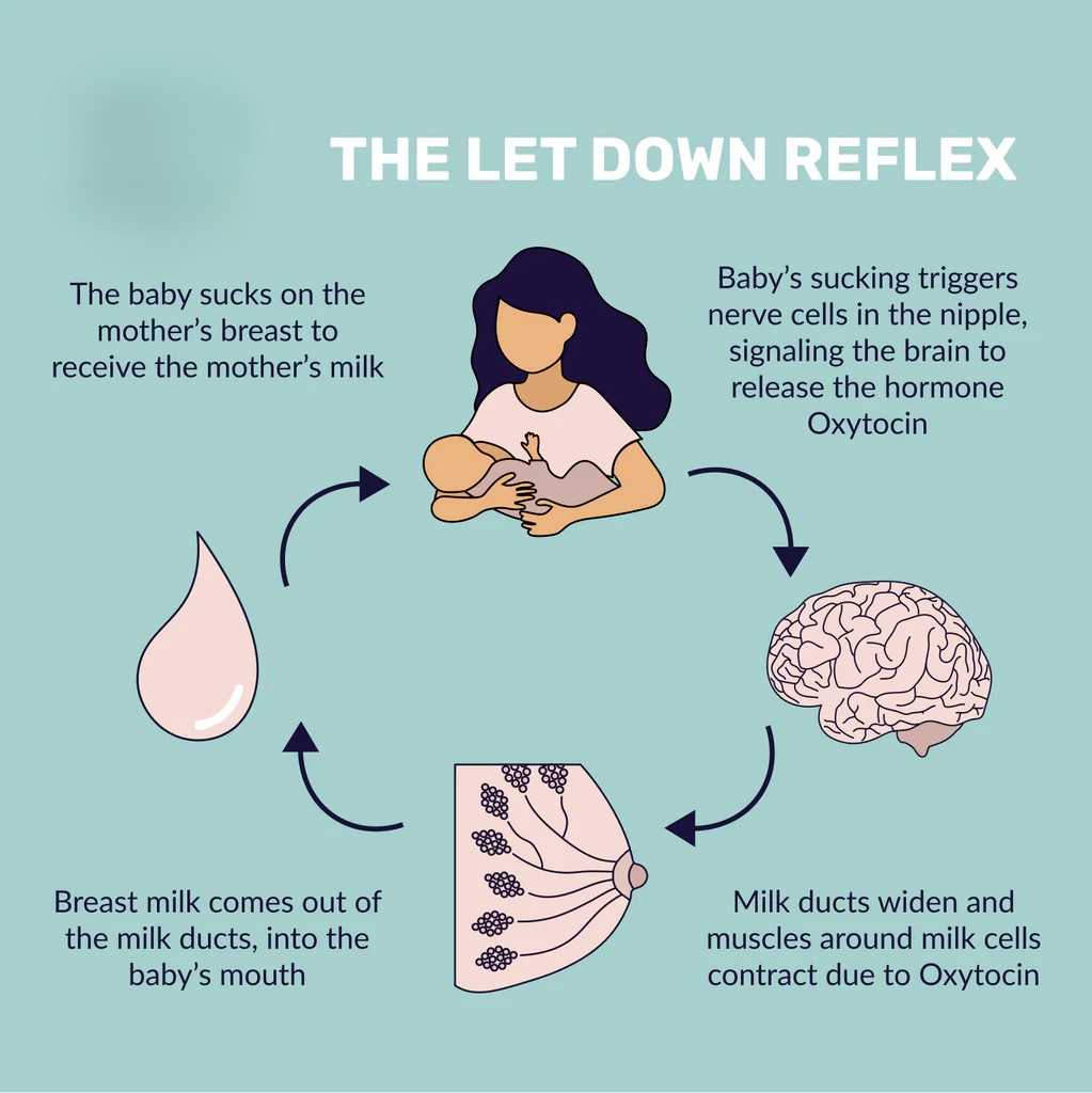
- Stimulates myoepithelial cell contraction in the breast, mediating milk ejection (let-down reflex) during lactation.
Hypothalamic Regulation and the Portal System
- Anterior pituitary relies exclusively on delivery of hypothalamic regulatory peptides via the hypophyseal portal venous system.
- Peptides are synthesized in hypothalamic nuclei, secreted into the primary capillary plexus at the median eminence, and travel down portal veins to the anterior lobe.
- Key Releasing Hormones:
- GHRH (Growth Hormone-Releasing Hormone): Stimulates GH.
- TRH (Thyrotropin-Releasing Hormone): Stimulates TSH and PRL.
- CRH (Corticotropin-Releasing Hormone): Stimulates ACTH.
- GnRH (Gonadotropin-Releasing Hormone): Stimulates FSH and LH.
- Key Inhibiting Hormones:
- Somatostatin (GHIH): Inhibits GH and TSH.
- Dopamine: Tonically inhibits PRL.
(Note: The integration of upstream hypothalamic signals dictates the ultimate secretory output of the anterior pituitary gland, whereas the posterior pituitary functions as a direct neural extension of the hypothalamus.)